Have you thought about what it would be like if early detection of breast cancer was more accessible and less invasive? What if you could monitor your breast health without having to book an annual mammogram? A team of researchers from MIT could make all of this possible.
Researchers have developed an innovative ultrasonic patch, designed to fit comfortably inside a bra, offering women a revolutionary way to monitor their health.
The need for early diagnosis
Breast cancer is a major health concern globally. Second the report “Global Cancer Statistics 2020”, new cancer cases in the world were approximately 19,3 million. Of these, female breast cancer accounted for 11,7% of new diagnoses, with 2,3 million of new cases estimated in 2020, making it the most frequently diagnosed cancer worldwide. Data that underlines the importance of research and development of new therapies to fight this disease.
The limits of traditional mammography
The most common method of testing for potential breast cancers is mammography, an X-ray image of the breast. Although mammography can detect lumps in breast tissue long before a doctor or individual can feel them, these screening methods they miss about one in eight breast cancers.
Then there is an insidious problem: from age 40 onwards, women are generally invited to undergo mammograms every year. However, for high-risk patients, this interval may not be sufficient. So-called “interval cancers,” which develop between routine scans, represent 20-30% of all breast cancer cases and they can be more aggressive.

The innovative solution from MIT
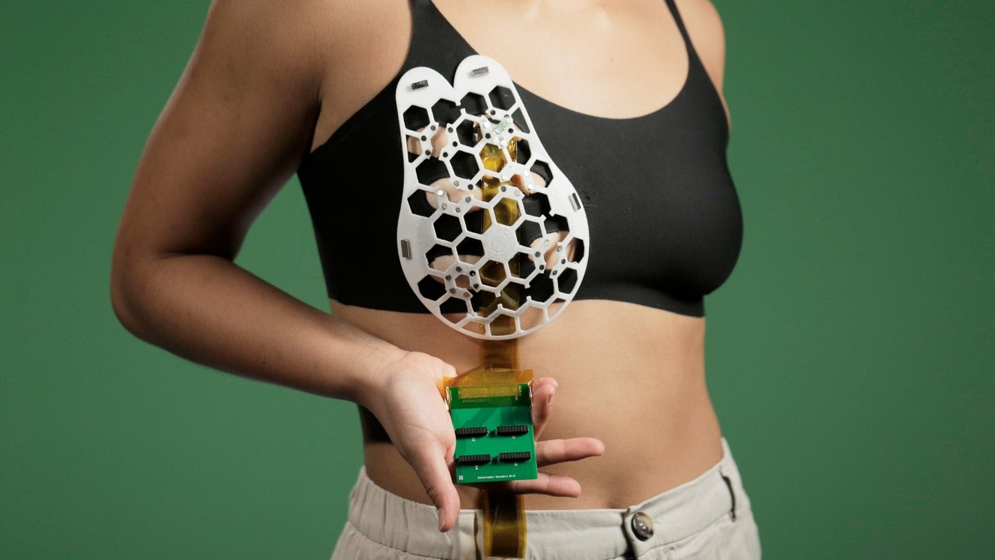
The flexible patch developed by MIT can obtain ultrasound images comparable to those made in medical centers, but it can be inserted into a bra. It's an amazing innovation, just featured in an article about Scientific Advances (that I link to you here).
“We have transformed ultrasound technology so it can be used at home. It is portable, easy to use and offers real-time, user-friendly monitoring of breast tissue,” declares Canan Dagdeviren, associate professor at MIT's Media Lab and senior author of the study.
Inspired by a true story
Moved by the story of her aunt, who died at 49 from breast cancer, Dagdeviren designed a small ultrasound scanner using piezoelectric material. This scanner can capture images whenever the user wants. The team also created a flexible 3D patch with “honeycomb” openings.
Paired with a bra, the scanner can be moved to six different positions to scan the entire breast, without the need for special training.
The wearable “mammography” device (the term is conventional, it is not X-rays obviously) was able to detect masses up to 8 centimeters deep and with a diameter of just 0,3 centimeters, maintaining a resolution similar to ultrasound traditional.

The future of diagnosis
In this stage of prototype development, users need to connect the diagnostic patch to a clinical-grade ultrasound device to view images. The team is already working on creating an imaging system the size of a phone.
In the future, high-risk people could use the device at home, repeatedly. Virtually even in real time. A decisive technology, especially for patients who do not have access to regular screenings.
An inclusive vision of health: from mammography to continuous screening
“Access to quality, affordable healthcare is essential for early diagnosis,” said the study author, Catherine Ricciardi, director of nursing at MIT's Center for Clinical and Translational Research.
“As a nurse, I have seen the negative consequences of a late diagnosis. This technology promises to break down barriers to diagnosis early breast cancer, offering a more reliable, comfortable and less intimidating diagnostic method.”
The future we want.