Researchers designed the 3D bioprinted model of anatomically accurate blood vessels, thus paving the way for possible advancements and new cardiovascular drugs.
Vascular diseases such as aneurysms and clots inside the blood vessels account for 31% of global deaths. Despite this, the progress of cardiovascular drugs have slowed down over the past 20 years. Why? Mainly, there is a lack of efficiency in converting possible treatments into approved methods. Particularly due to the discrepancy between studies that occur on the outside of the body versus the inside.
Recent research aims to reshape current methodologies to minimize this gap and improve the translatability of these techniques by directing 3D bioprinting towards vascular medicine. This interdisciplinary and collaborative project was recently published in the Advanced Healthcare Materials journal.
Printing “perfect” blood vessels in 3D
3D bioprinting is an advanced manufacturing technique capable of producing unique tissue-shaped constructs with embedded cells layer by layer, making the arrangement more likely to mirror the native multicellular composition of blood vessels. A range of hydrogel bioinks have been introduced to engineer these structures: However, there is a limitation in available bioinks that can mimic the vascular composition of native tissues. Current bioinks lack high printability and are unable to deposit a high density of living cells in complex 3D architectures, making the process less effective.

To overcome these shortcomings, Dr. Akhilesh Gaharwar and the professor Abhishek jain Texas A&M have developed a new nano-engineered bio-ink to print 3D, anatomically accurate, multicellular blood vessels. Their approach offers improved real-time resolution for both macrostructure and tissue-level microstructure.
And this was not possible with the available bio-inks.
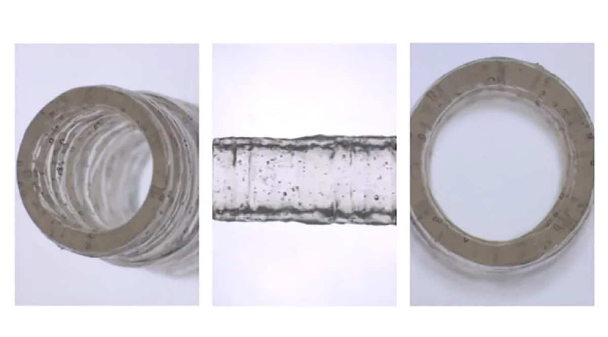
A surprising bio-ink
“A remarkably unique feature of this nanoengineered bioink is that, regardless of cell density, it demonstrates high printability. It has the ability to protect the encapsulated cells from high shear forces in the bioprinting process,” says Gaharwar. “Surprisingly, the 3D bioprinted cells maintain a healthy phenotype and remain viable for almost a month after fabrication.”
Taking advantage of these unique properties, the nanoengineered bioink is printed into 3D cylindrical blood vessels, consisting of living co-cultures of endothelial cells and vascular smooth muscle cells. This will give researchers the opportunity to model vascular function and disease impact.