The battle against cancer is enriched with a new milestone, and it's all 3D printed.
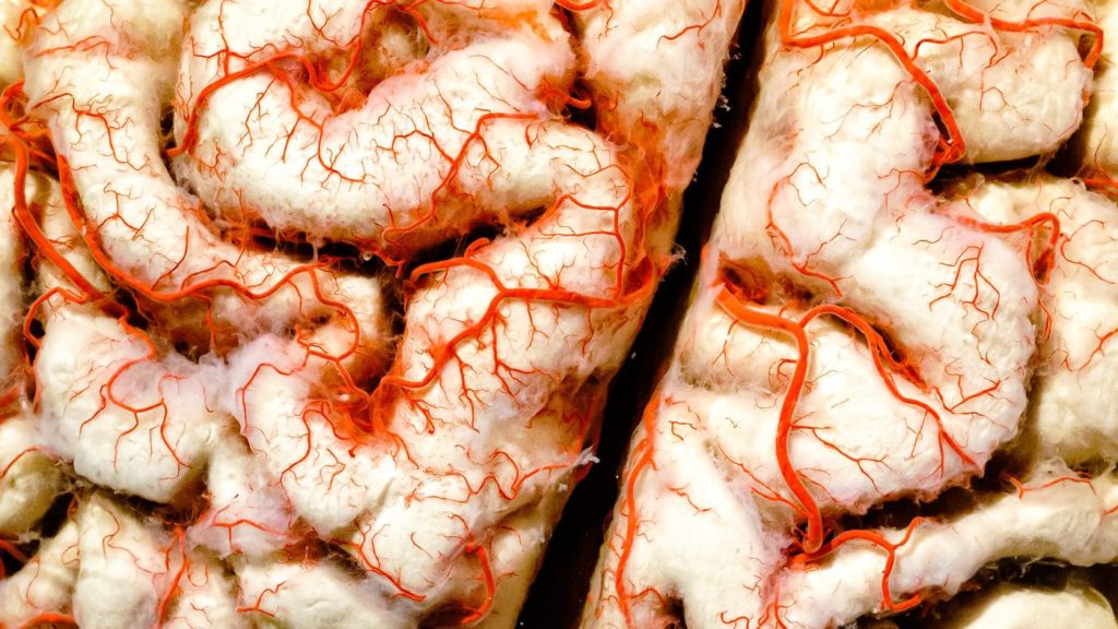
Researchers from Tel Aviv University (TAU) they printed in 3D a brain tumor. It is not something far from the real model: it is a real active glioblastoma. It is one of a kind, in a brain-like environment, complete with blood vessels that supply the mass.
3D printing a brain tumor could pave the way for the development of new methods (and research in simulated environments). Methods to improve treatment and accelerate the discovery of new drugs.
The most accurate replica ever of a brain tumor
According to the researchers, this is the largest replication of a brain tumor and surrounding tissue to date. The 3D model created includes “a complex system of tubes similar to blood vessels. Blood cells and drugs can flow through them, simulating a real brain tumor."
The extraordinary study was published in the journal Science Advances.
Glioblastoma and the breakthrough
Glioblastoma is an aggressive type of cancer that can form in the brain or spinal cord, and although it can be rare, it is particularly frightening as it develops rapidly and is almost always fatal. All of this makes treatment extremely difficult, which is why therapy must be rigorous, with courses of chemotherapy and radiotherapy that patients often cannot even complete.
New drugs could always help. However, current drug development processes are time-consuming and do not show how a drug will act in a patient's body.
“Cancer, like all tissues, behaves very differently in a Petri dish or a test tube than in the human body,” says lead researcher Prof. Ronit Satchi-Fainaro in a release. "About 90% of all investigational drugs fail in clinical trials because the success obtained in the laboratory is not reproduced in patients."
3D printing of a brain tumor
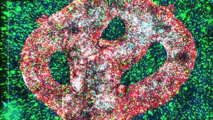
To try to overcome these problems, TAU scientists turned to 3D printing. Through rigorous research, they were able to create the world's first fully operational 3D model of a brain tumor such as glioblastoma, complete with 3D printed cancerous tissue. and the surrounding tumor environment that influences its development.
The “printed” tumor is made of a gel composition of a similar consistency to the brain and features a sophisticated system of blood vessel-like tubes through which blood cells and drugs can flow. This allows researchers to see how a real tumor forms and responds to treatments.
How will the treatment protocol be
“The process in which we bioprint a tumor from a patient is that you go into the operating room, extract tissue from the tumor, and print it based on that patient's MRI,” explains Satchi-Fainaro. “So, we have about two weeks where we can test all the different therapies to evaluate their effectiveness for that specific brain tumor and come back with an answer as to which treatment is expected to be the most suitable.”
100 tumors, 100 attempts
“If we take a sample from a patient's tumor, together with the surrounding tissues, we can 3D bioprint 100 tiny tumors from this sample and test many different drugs in various combinations to discover the optimal treatment for this specific tumor,” says the researcher.
“Alternatively, we can test numerous compounds on a 3D bioprinted tumor and decide which is most promising for further development and investment as a potential drug.”
The researchers were able to use their new technique to target a specific protein pathway that allows the immune system to help the brain tumor spread rather than kill fatal cancer cells. As a result, the growth of the glioblastoma was slowed and the invasion was stopped.
We have demonstrated that our 3D printed model is best suited for predicting treatment efficacy, drug target discovery, and new drug development
Ronit Satchi-Fainaro