

A statement from the Great Ormond Street Hospital (GOSH) for Children in London suggests that the baby with spina bifida directly in the womb is healthy and developing well.
What is spina bifida
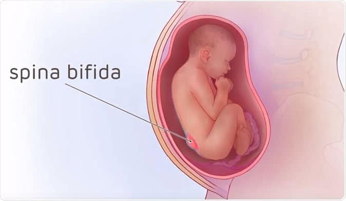
Occurs when the so-called neural tube, a hollow structure that begins to form around the third week of pregnancy, does not develop properly and essentially ends up with a hole. The neural tube will later give rise to the baby's brain and spinal cord, which is why a hole in the structure can cause mild to severe nerve damage and result in physical and intellectual disabilities.
Helena (not her real name), a mother in the UK, learned that her baby had spina bifida during her fifth month of pregnancy.
“It was a very large lesion on her back and half her spine was exposed,” Helena told BBC News.
They said it was likely she was paralyzed, incontinent, and would later need a shunt to drain fluid from her brain.
A hope from the last decade
Luckily, in 2011, a landmark clinical trial showed that operating on babies in the womb could spare them from some of the harmful effects of spina bifida. Until 2019, however, it was an unbeaten approach.
Compared with children with spina bifida who had surgery after birth, those operated on in utero were twice as likely to walk unassisted at age 2 and with fewer neurological problems.
Fetal surgery involves some risks, especially those of premature birth and requires a cesarean section.
“The spina bifida procedure is complex and not without risks, but it can change the lives of children and their families. It cannot be overstated,” says Dr. Dominic Thompson, chief neurosurgeon at the English facility.
A collective effort
Helen's operation involved 25 doctors from GOSH and University College London Hospitals, as well as University Hospitals Leuven in Belgium, where the operation was physically conducted.
In general, the procedure involves first administering an anesthetic to the mother, which is also passed on to the fetus. The spinal column of the fetus is then reached through the mother's abdomen and uterus. Here comes the trickiest step: Neurosurgeons separate any skin attached to the exposed spinal cord and place the cord inside the spinal canal before stitching the tissues closed.
Helena received a surgery in her 23rd week of pregnancy and three months later gave birth to her daughter Mila at University College London Hospital. There's still some excess fluid in the little girl's brain, but so far Mila is showing signs of healthy development.
“He can move his legs and has feeling in his toes, it's absolutely amazing,” his mother says. “I'm just so grateful to the surgeons who did this operation because her life would be so different without it.”
With Mila, the team has performed the same operation on 32 children since January 2020.
Spina bifida: there's also a bit of Italy
“We are very excited about the next phase of prenatal surgery for children with spina bifida, including less invasive approaches,” says Dr. Paul DeCoppi, Italian doctor and surgeon who is part of the fetal surgical team for spina bifida.
As with any new approach, the benefits and risks for the mother and baby must now be fully understood. What is clear, however, is that prenatal surgery for patients with spina bifida leads to better results.

